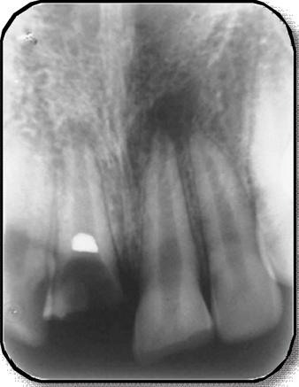
A) Preoperative intraoral periapical (IOPA) radiograph of 36. B
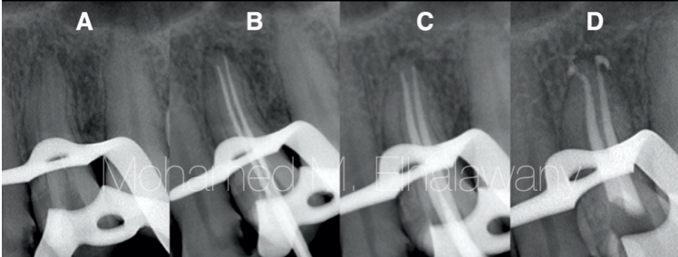
A) Preoperative intraoral periapical (IOPA) radiograph of 36. B) Post operative (IOPA) radiograph of 36. C) 1 month follow up IOPA radiograph of 36. D) 6 months follow up IOPA radiograph of 36. E) 1 year follow up IOPA radiograph of 36. - IP Indian J Conserv Endod - clinical and preclinical conservative /restorative de

Radiographic aids in dx of periodontol ds

a, b) Preoperative view of #36 and #46. (c, d) Preoperative IOPA

a) Preoperative IOPA radiograph of tooth #36. (b) Intraoral image

Direct pulp capping with bioactive materials – A case series - IJCE

Deepshikha CHOWDHURY, Master of Dental Surgery

Apexogenesis of immature permanent molar using MTA with long term
jcdr-11-ZD05-g003.jpg

Tooth 36. (A) The preoperative periapical radiograph. (B) The

Endodontic Management of the Maxillary First Molar with Two

Paromita MAZUMDAR, Head of Faculty

Radiographic aids in dx of periodontol ds

in Endodontics Pocket Dentistry
Pre-operative X-ray: suggested or obligatory - Style Italiano

a) Preoperative IOPA radiograph of tooth #36. (b) Intraoral image