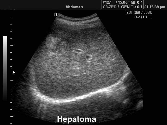
Ultrasound images • Hepatoma, B-mode, echogramm №103
Abdomen: Liver (hepatoma) in B-mode. Ultrasound image №103 was received by the scanner SonoAce-8000.
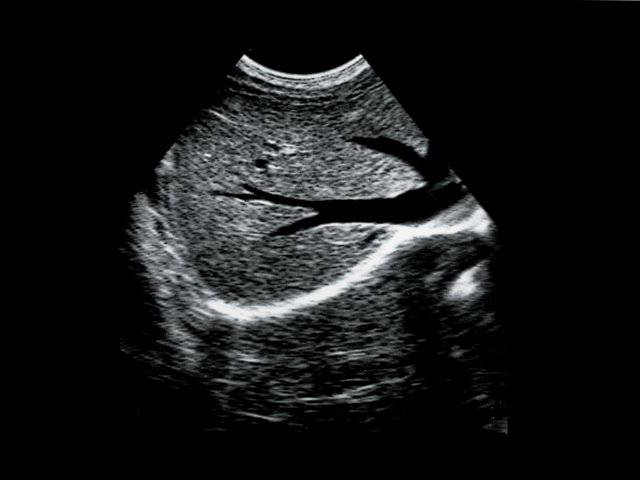
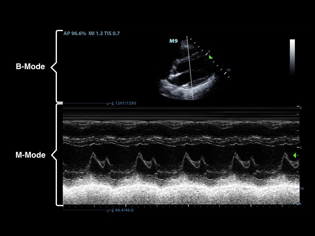
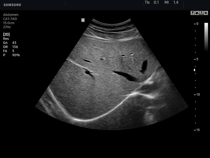
Ultrasound images • Liver and hepatic veins, B-mode, echogramm №45
Cancers, Free Full-Text

Sensors, Free Full-Text

EPOS™

Sensors, Free Full-Text
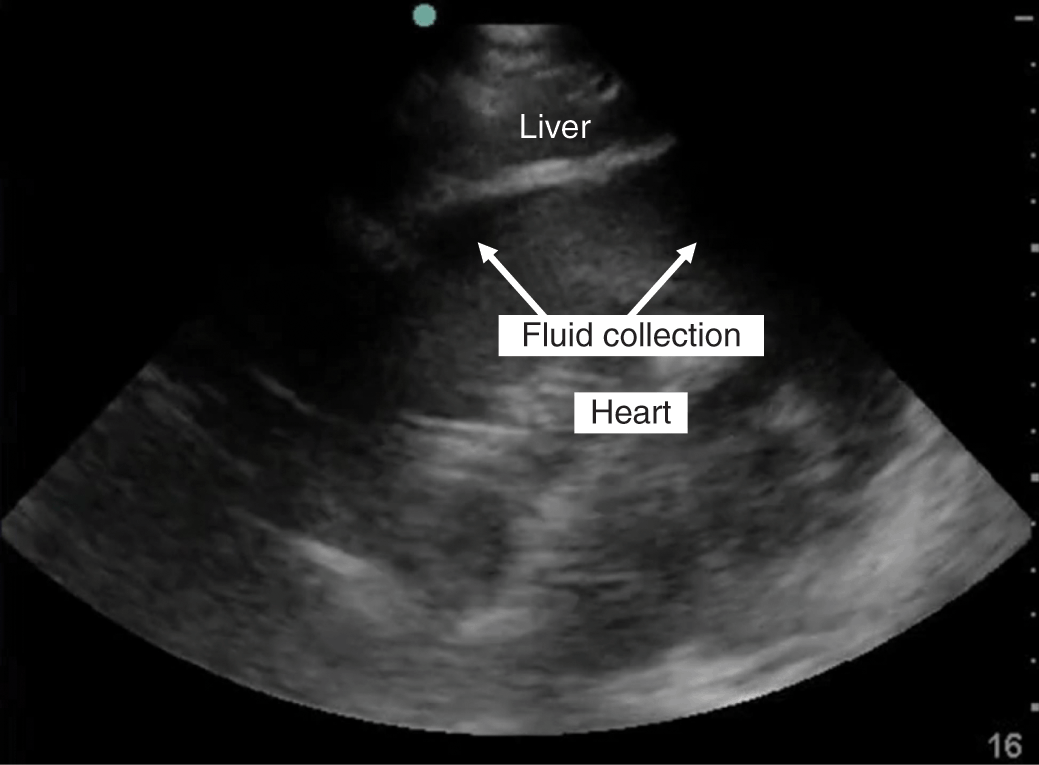
Ultrasound in Trauma (Chapter 25) - Color Atlas of Emergency Trauma
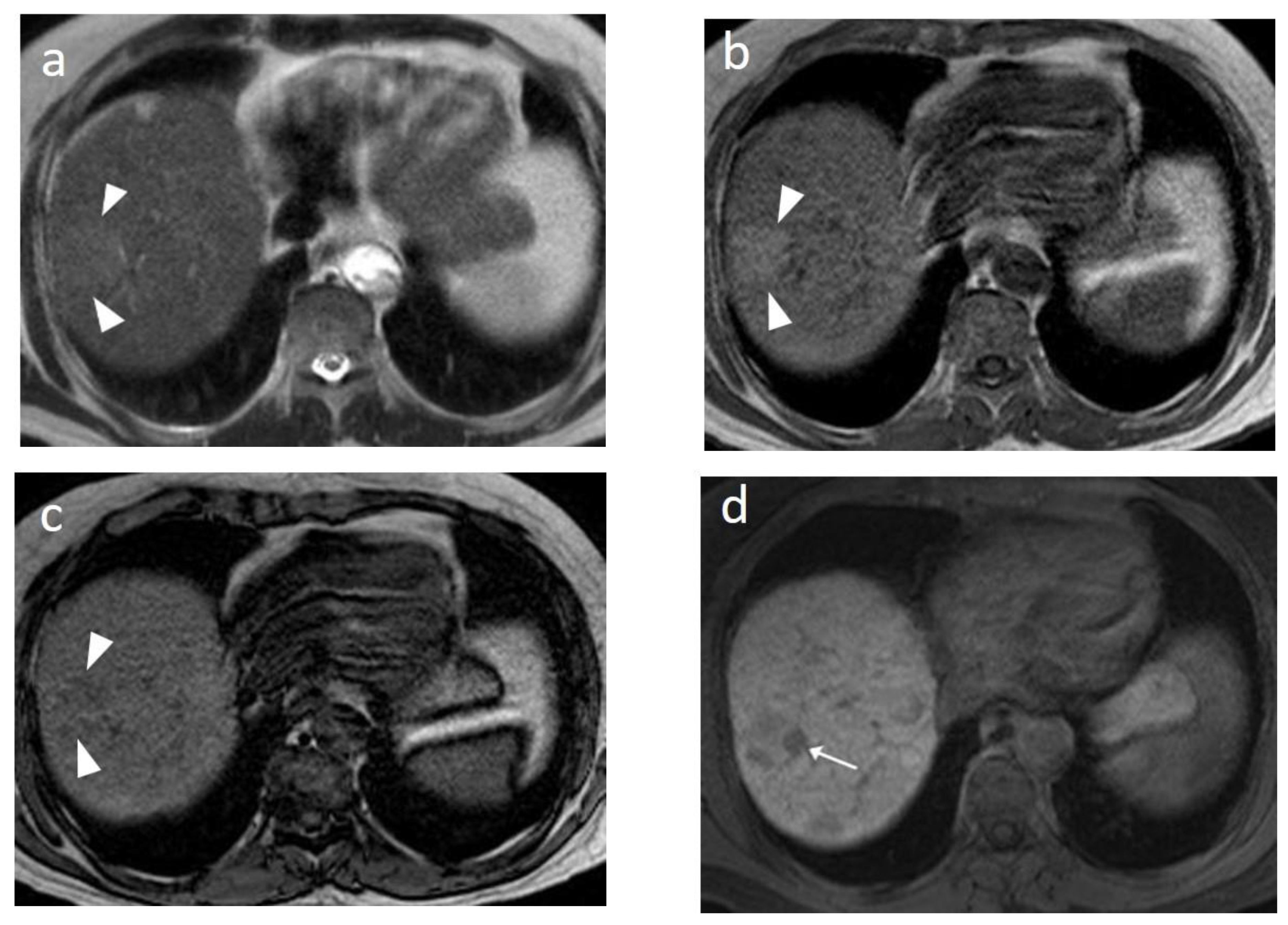
Transabdominal ultrasound performed at 27 GW. (a) Hepatomegaly, liver
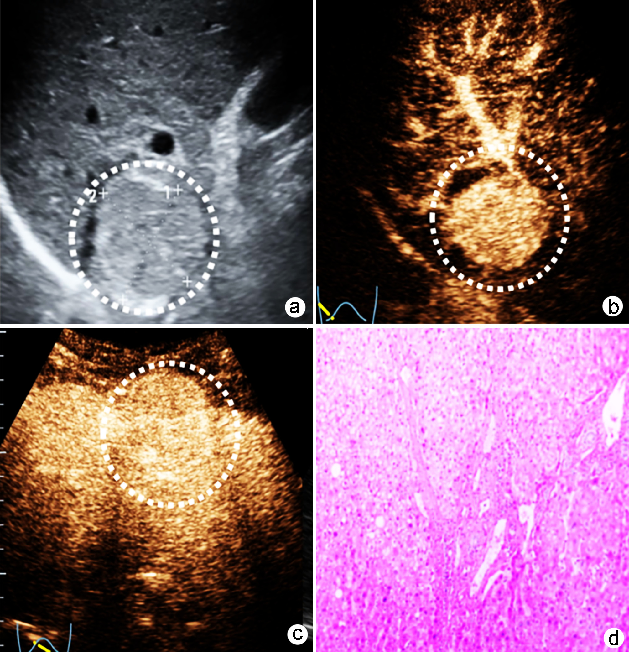
Construction and validation of a nomogram model for microvascular invasion in hepatocellular carcinoma based on the characteristics on contrast-enhanced ultrasound Liver Imaging Reporting and Data System
Sensors, Free Full-Text
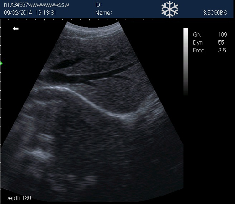
Ultrasound images • Liver, B-mode, echogramm №742

Total abdominal ultrasound shows: (A) and (B) hepatomegaly secondary
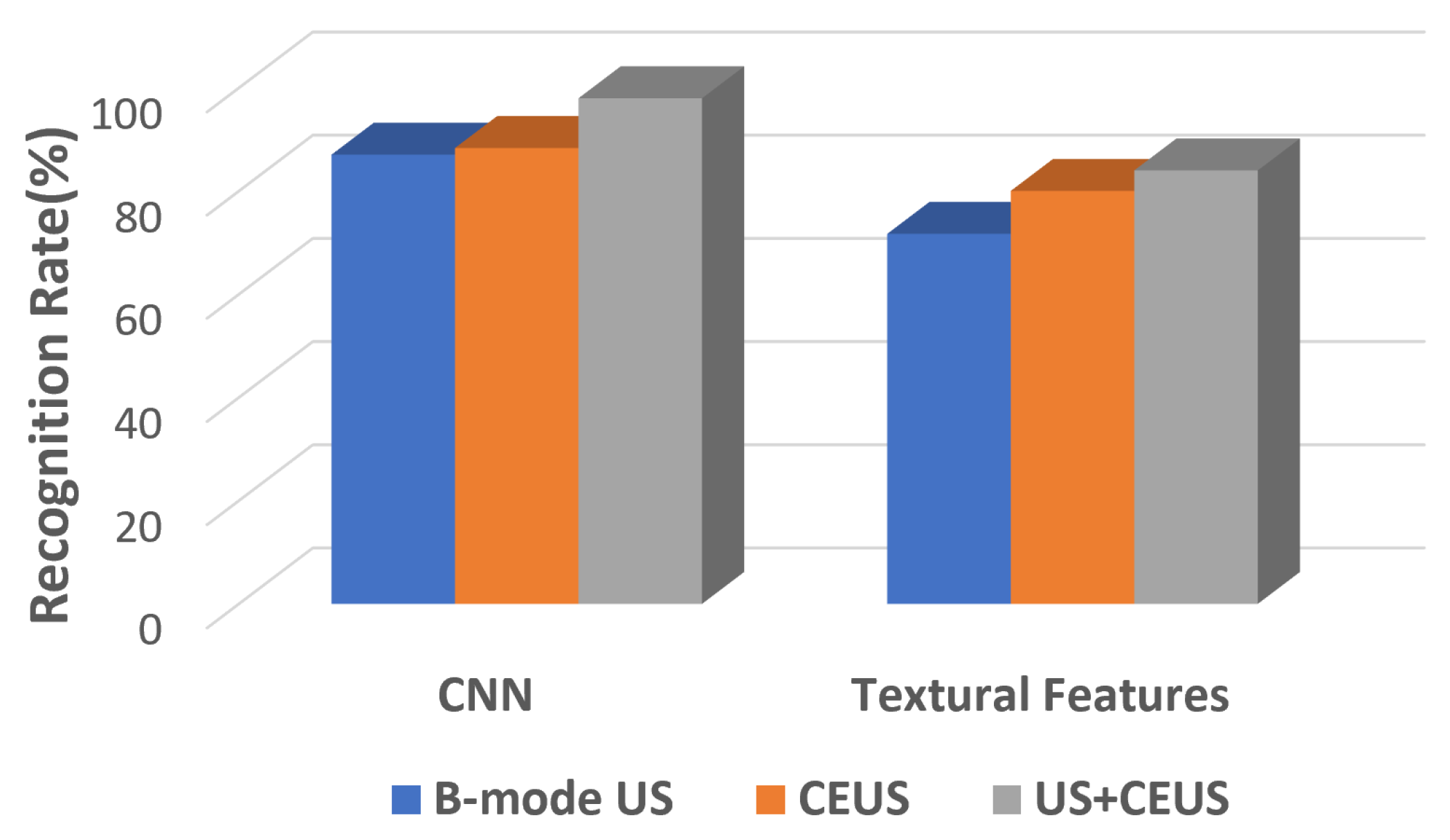
Sensors, Free Full-Text
Hepatoblastoma, Radiology Reference Article

Color Duplex Scanning of the Hepatoportal Circulation

Profiling hepatocellular carcinoma aggressiveness with contrast-enhanced ultrasound and gadoxetate disodium-enhanced MRI: An intra-individual comparative study based on the Liver Imaging Reporting and Data System - ScienceDirect