Iliotibial Band Friction Syndrome - MSK Radiology Imaging
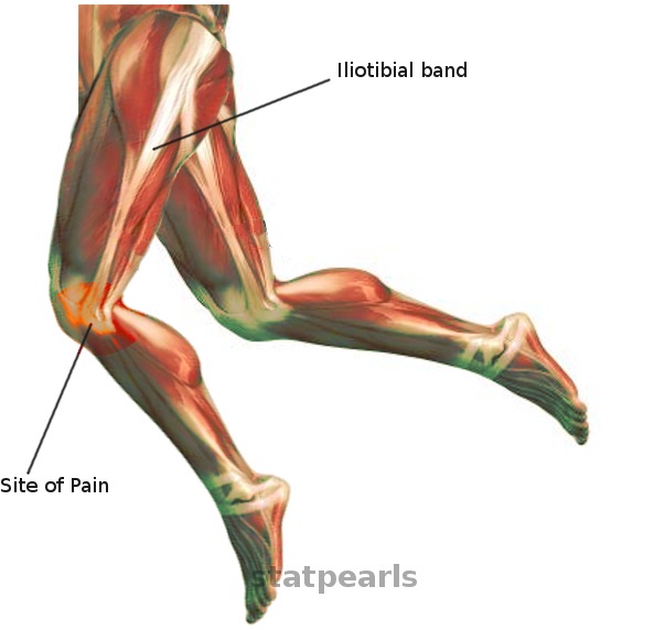
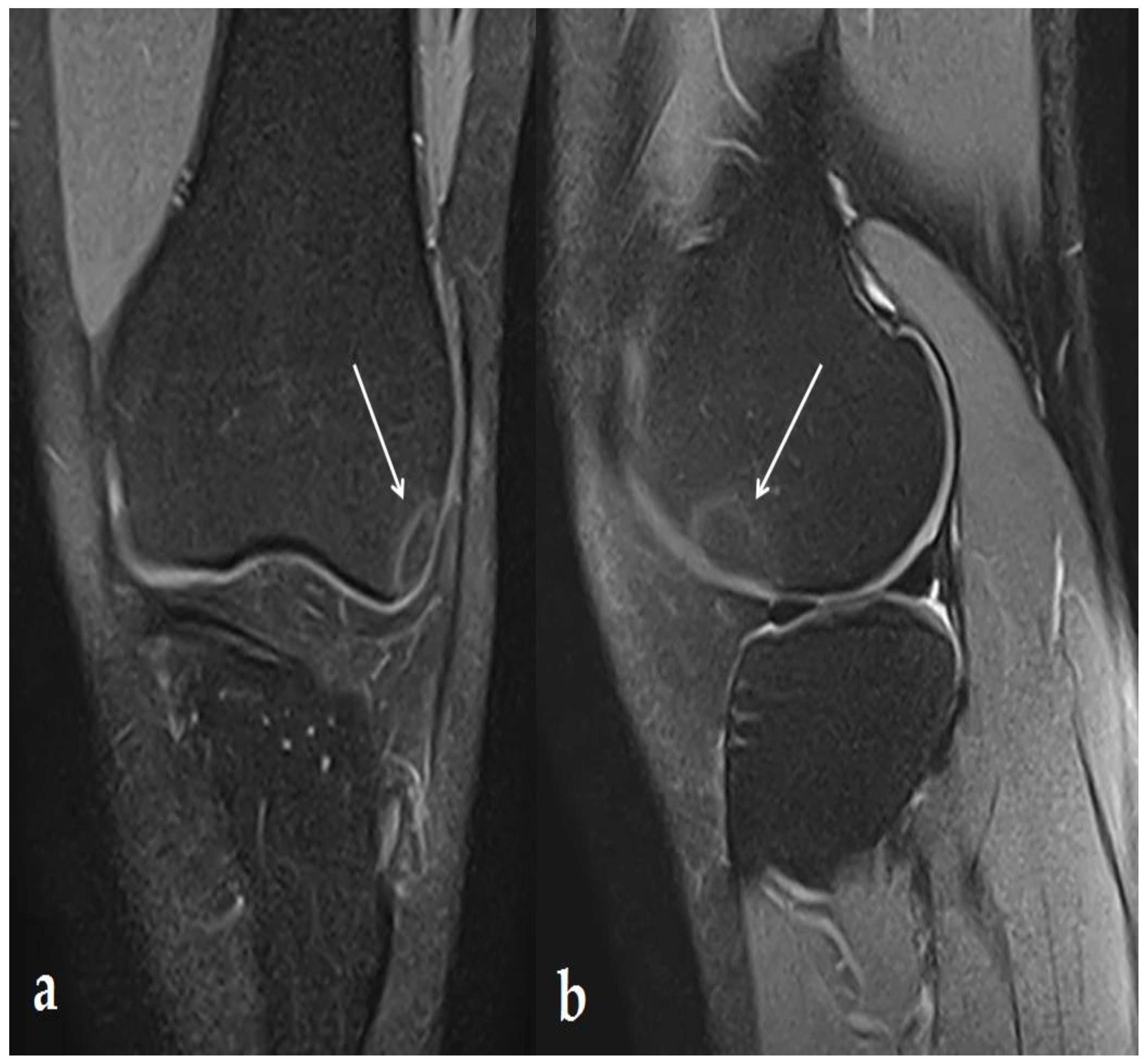
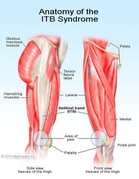
Iliotibial Band Friction Syndrome - MSK Radiology Imaging Findings: • The iliotibial band (ITB) is thickened with no tear visualized. • Ill-defined area of increased signal on fluid-sensitive sequences between the lateral femoral condyle (LFC) and ITB. Case description: • Clinical: anterolateral knee pain with point tenderness 1-2 cm proximal to lateral joint line. • Treatment: Conservative measures and image-guided steroid injection (may accelerate recovery). • Chronic inflammatory response to friction between the TIB and LFC causing ill-defined increased signal in this region on fluid-sensitive sequences. • Findings of chronic disease: - Thickening of the IT B and superficial increased T2-signal. - Reactive marrow edema in the adjacent LFC. Differential diagnosis for similar location of pain: • Fluid in lateral knee joint recess: Well-defined margins and connection to knee joint is seen. • Lateral collateral ligament complex injury: Signal around and/or within lateral ligaments. • Direct trauma/contusion: Soft-tissue swelling is predominant, with minimal fluid-signal deep to ITB. Dr. Donald von Borstel @DrvonBorstel #Iliotibial #Band #ITBand #Friction #Syndrome #Radiology #diagnosis #msk #clinical

Magnetic Resonance Imaging of Iliotibial Band Syndrome

ultrasound Guided Interventions: Healthy Images (Iliotibial band)
IJERPH, Free Full-Text

ITB ILIOTIBIAL BAND FRICTION SYNDROME MRI: IS IT A BURSA OR A RECESS? - Radedasia

Iliotibial Band Syndrome (ITBS) Southern California Orthopedic Institute

Iliotibial Band Syndrome (“Runner's Knee”)

Iliotibial band syndrome, Radiology Case
Treatment of ITB Syndrome at Our Clinic - New York Dynamic Neuromuscular Rehabilitation

Iliotibial band friction syndrome Cortisone injection

Imaging of knee by mr and usg

ITB ILIOTIBIAL BAND FRICTION SYNDROME MRI: IS IT A BURSA OR A RECESS? - Radedasia