![Figure, B-Mode ultrasound showing main portal] - StatPearls](https://www.ncbi.nlm.nih.gov/books/NBK567725/bin/pv.jpg)
Figure, B-Mode ultrasound showing main portal] - StatPearls
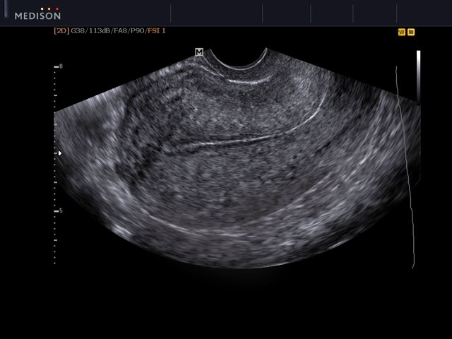
B-Mode ultrasound showing main portal vein diameter of 15.1 millimeters. This is an indirect finding of portal hypertension. Contributed by Brian Covello, MD

Sonography of a Typical Parathyroid Adenoma: Solitary Parathyroids as Seen on Ultrasound
Salivary gland ultrasound in primary Sjögren's syndrome

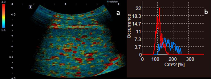
Dual imaging with standard B-mode sonography (a) and contrast enhanced

Invasive mole, Radiology Case
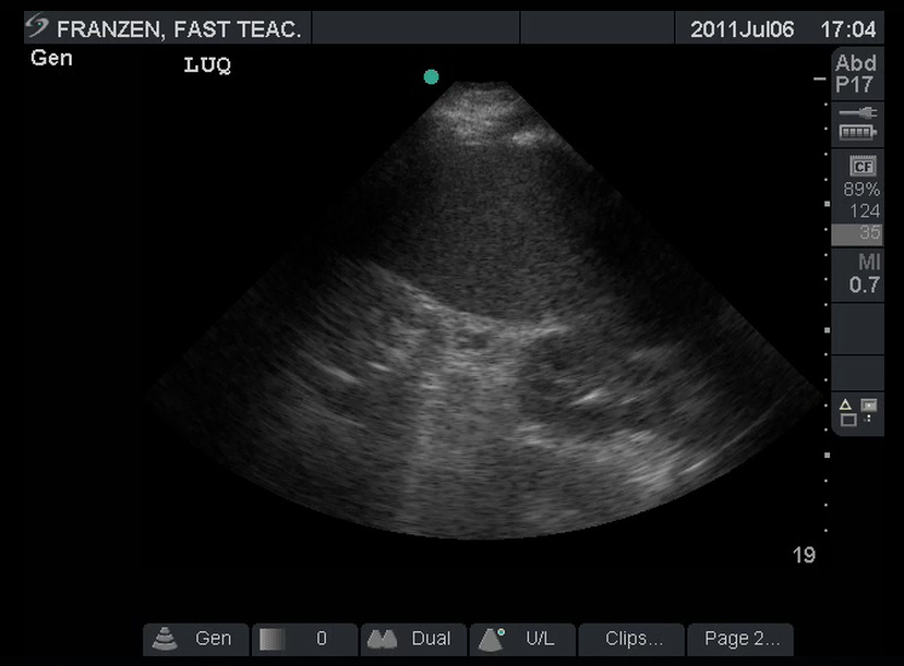
FAST Exam

Invasive mole, Radiology Case

Ultrasound B-mode examination of the 4-chamber (A, B) and 3-vessel (C

B-mode image of the spleen constructed by ultrasound-fusion image. The

– Emergency Medicine EducationSplenic Infarction: ED Presentation, Evaluation, and Management - - Emergency Medicine Education

Operator Evaluation of Ultrasound Fusion Imaging Usefulness in the Percutaneous Ablation of Hepatic Malignancies: A Prospective Study - ScienceDirect
The severity of portal hypertension by a non-invasive assessment: acoustic structure quantification analysis of liver parenchyma, BMC Medical Imaging