Optical Coherence Tomography: Imaging Mouse Retinal Ganglion Cells In Vivo
Scientific Article | Structural changes in the retina are common manifestations of ophthalmic diseases.
Quantification of optical In-Vivo imaging of retinal and choroidal neovascularisation and cell migration in the mouse fundus - MedCrave online

Optical Coherence Tomography: Imaging Mouse Retinal Ganglion Cells In Vivo
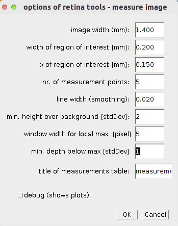
Retina Tool - ImageJ-macros - MRI's Redmine

All Protocols and Video Articles in JoVE

SM-OCT imaging of the mouse cornea and retina clarifies the
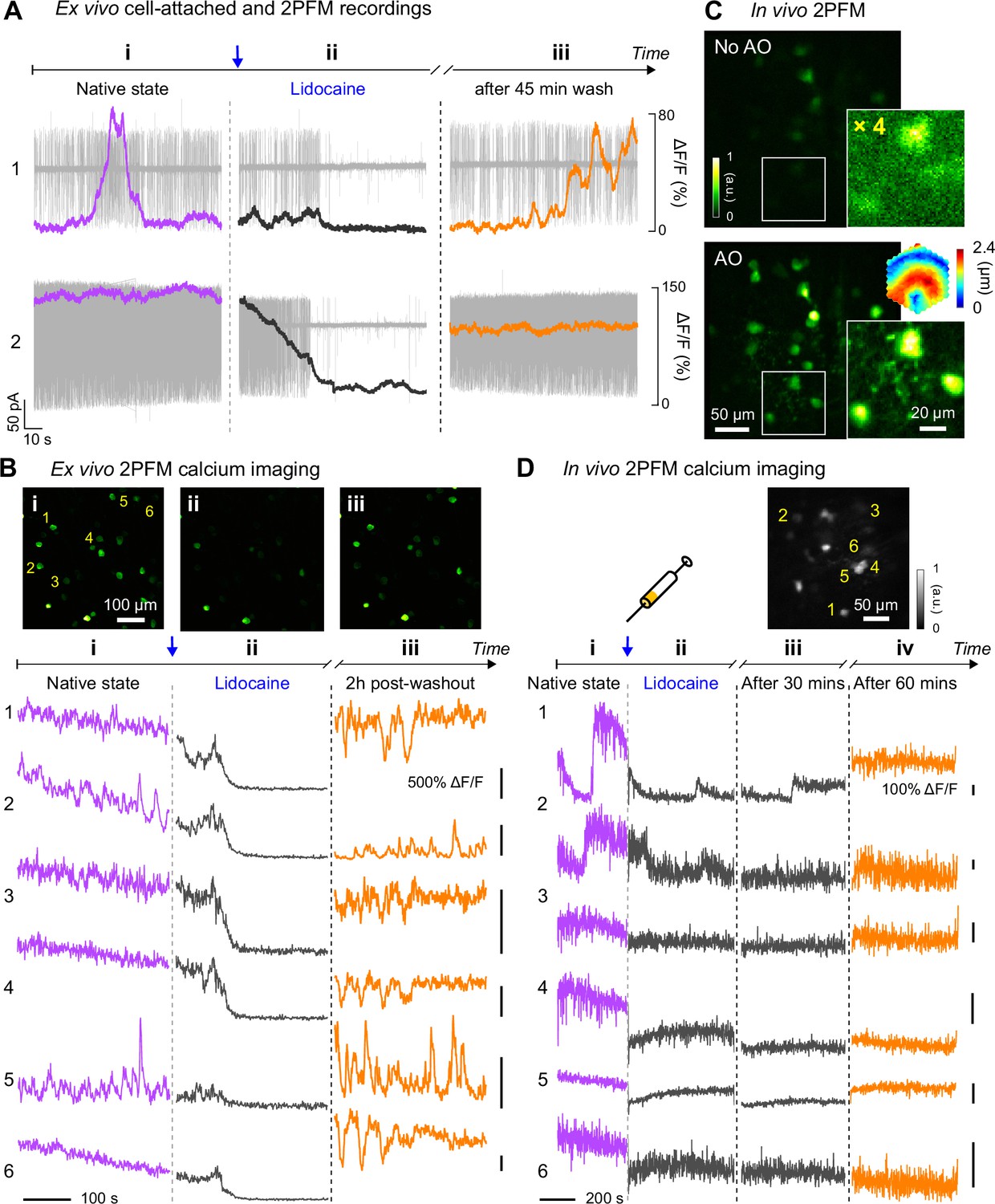
Retinal microvascular and neuronal pathologies probed in vivo by adaptive optical two-photon fluorescence microscopy
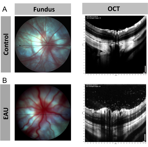
Image-Guided Optical Coherence Tomography to Assess Structural Changes in Rodent Retinas

All Protocols and Video Articles in JoVE
Aplicação da Tomografia de Coerência Óptica a um Modelo de Retinopatia de Rato

Emmanuelle SARZI, Professor (Assistant), Claude Bernard University Lyon 1, Villeurbanne, UCBL, Institut NeuroMyogène

OCT imaging and phase-variance (pv-) analysis reveals the mouse retinal

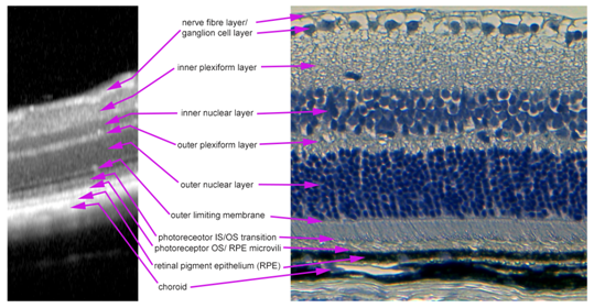
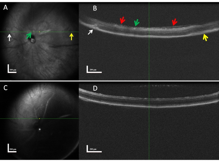
OCT imaging of morphology in mouse posterior eye. A) OCT fundus image
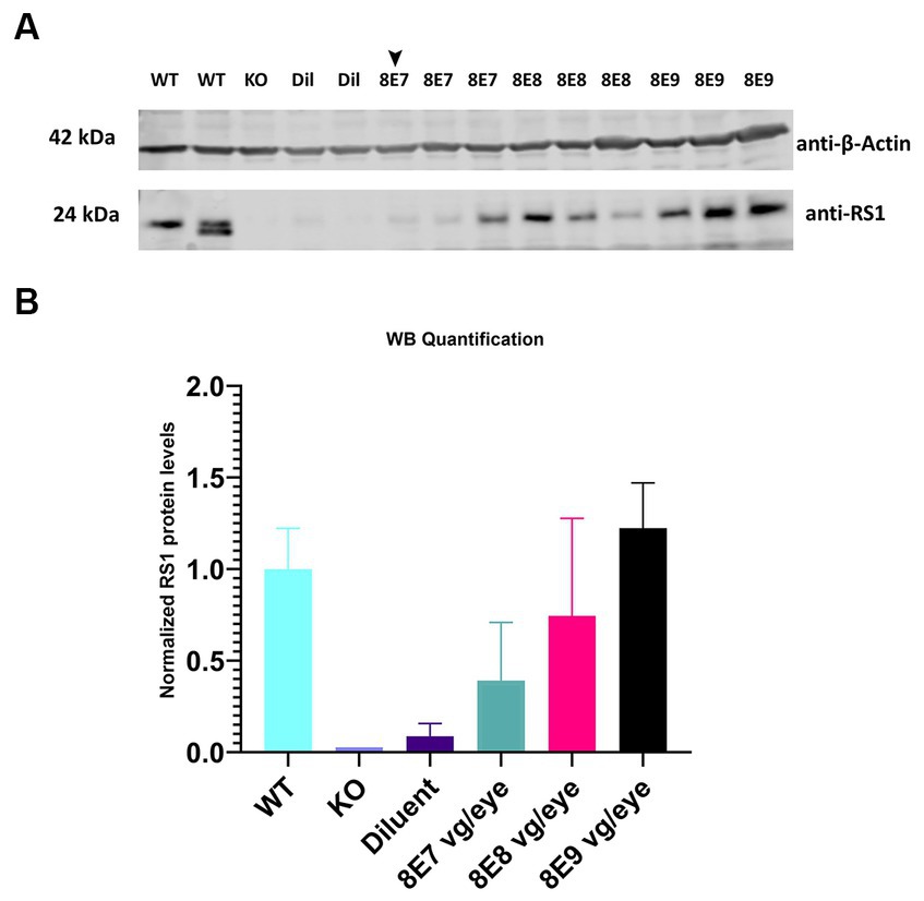
Frontiers The dose-response relationship of subretinal gene therapy with rAAV2tYF-CB-hRS1 in a mouse model of X-linked retinoschisis