![PDF] Dry Socket Etiology, Diagnosis, and Clinical Treatment Techniques](https://d3i71xaburhd42.cloudfront.net/c80eaa81ee0ce272f24e5fe84be66723e78371ff/2-Figure1-1.png)
PDF] Dry Socket Etiology, Diagnosis, and Clinical Treatment Techniques
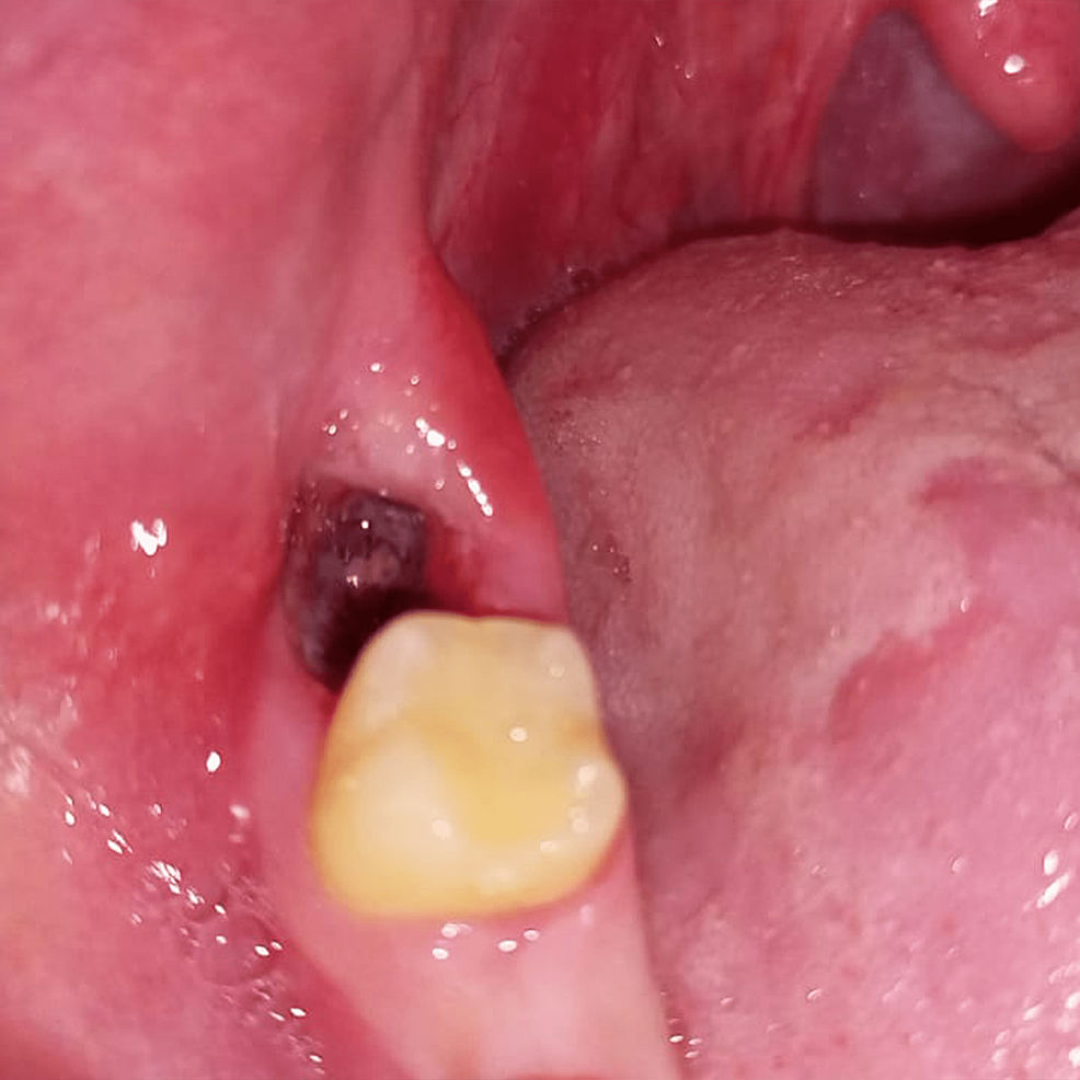
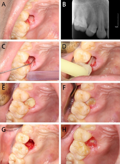
How microscope level loupe magnification of 6× to 8× or greater, combined with co-axial illumination or a dental operating microscope, facilitate more precise treatment of dry socket lesions is shown. Dry socket, also termed fibrinolytic osteitis or alveolar osteitis, is a complication of tooth exodontia. A dry socket lesion is a post-extraction socket that exhibits exposed bone that is not covered by a blood clot or healing epithelium and exists inside or around the perimeter of the socket or alveolus for days after the extraction procedure. This article describes dry socket lesions; reviews the basic clinical techniques of treating different manifestations of dry socket lesions; and shows how microscope level loupe magnification of 6× to 8× or greater, combined with co-axial illumination or a dental operating microscope, facilitate more precise treatment of dry socket lesions. The author examines the scientific validity of the proposed causes of dry socket lesions (such as bacteria, inflammation, fibrinolysis, or traumatic extractions) and the scientific validity of different terminologies used to describe dry socket lesions. This article also presents an alternative model of what causes dry socket lesions, based on evidence from dental literature. Although the clinical techniques for treating dry socket lesions seem empirically correct, more evidence is required to determine the causes of dry socket lesions.
The efficacy of minocycline hydrochloride ointment versus iodoform

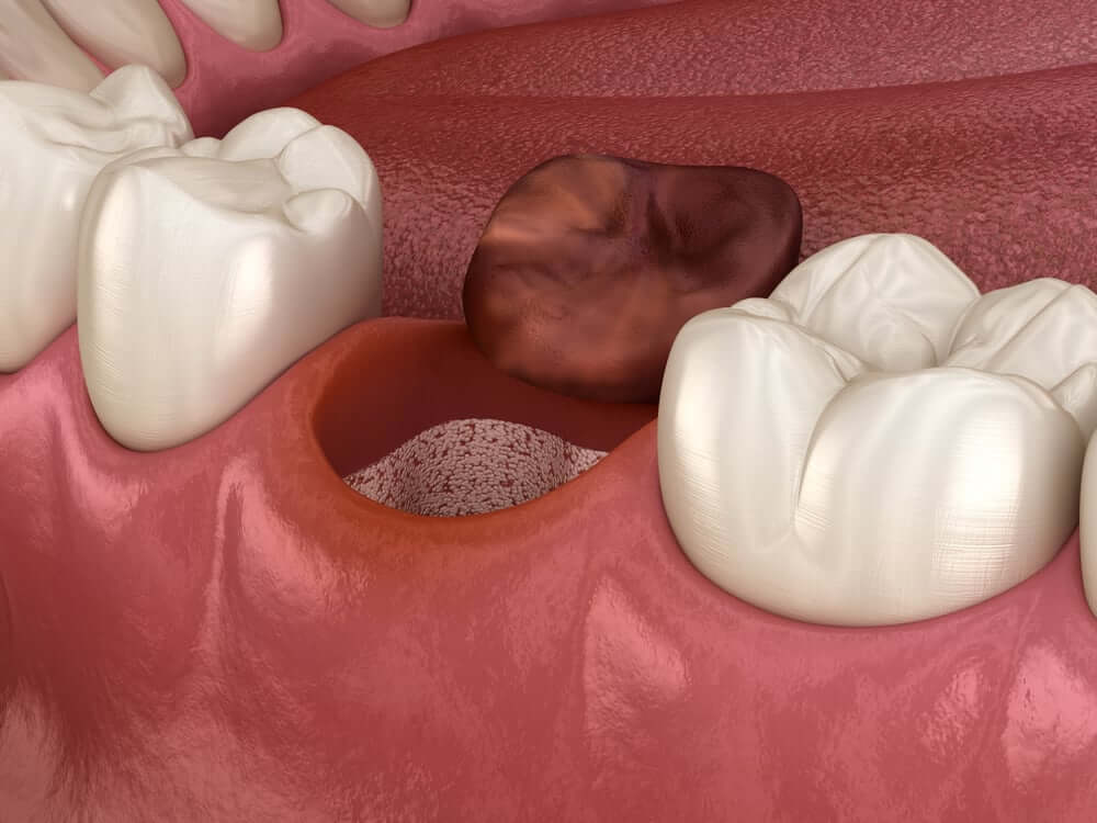
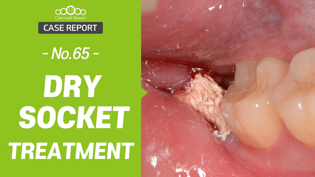
Dry socket
Applied Sciences, Free Full-Text

9 Home Remedies For Dry Socket And Prevention Tips
PDF) Curettage versus Zinc Oxide Eugenol as a Treatment Modality
PDF) Painful dry socket: an alternative perspective

Dry socket

The non-healing extraction socket: a diagnostic dilemma - case

Dry socket