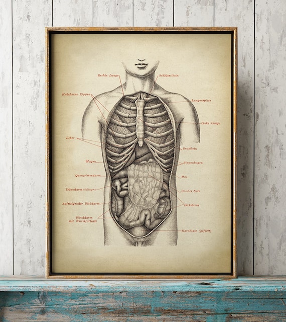
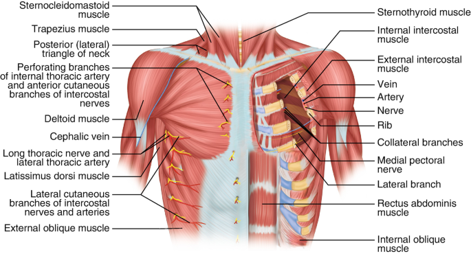
Figure 3 from Relevant surgical anatomy of the chest wall.
Fig. 3. Anterior chest wall showing the sternum. Note where the costal cartilages articulate with the sternum. In the intercostal space lie different structures: several kinds of intercostal muscles, intercostal arteries and associated veins, lymphatics, and nerves. (From Rendina EA, Ciccone AM. The intercostal space. Thorac Surg Clin 2007;17(4):491e501; with permission.) - "Relevant surgical anatomy of the chest wall."

Heart Anatomy Anatomy and Physiology II

Bones and joints of the thoracic wall: Video

Figure 3 from Relevant surgical anatomy of the chest wall.
Surgeries, Free Full-Text

Surgical Anatomy of the Chest Wall

Introduction to chest wall reconstruction: anatomy and physiology of the chest and indications for chest wall reconstruction. - Abstract - Europe PMC
Chest Wall Anatomy
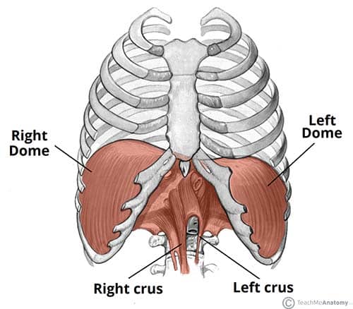
The Diaphragm - Actions - Innervation - TeachMeAnatomy
Chest Wall Tumour: Causes, Symptoms, Signs, and Treatment - MyHealth

Minimally Invasive Surgical Correction of Chest Wall Deformities in Children (Nuss Procedure) - Advances in Pediatrics

pub.mdpi-res.com/jcm/jcm-11-05516/article_deploy/h