This scanning electron micrograph (SEM) depicted a number of red
Download this stock image: This scanning electron micrograph (SEM) depicted a number of red blood cells found enmeshed in a fibrinous matrix on the luminal surface of an indwelling vascular catheter; Magnified 11432x Note the biconcave cytomorphologic shape of each erythrocyte, which increases the surface area of these hemoglobin-filled cells, thereby, promoting a greater degree of gas exchange, which is their primary function in an in vivo setting. In their adult phase, these cells possess no nucleus. What appears to be irregularly-shaped chunks of debris, are actually fibrin clumps, which when inside the living organi - 2BE0H0B from Alamy's library of millions of high resolution stock photos, illustrations and vectors.

A. Scanning electron microscope picture of red blood cells subjected
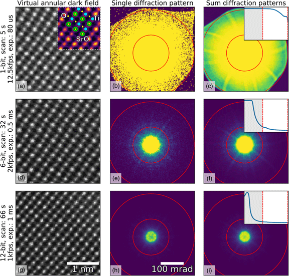
Fast Pixelated Detectors in Scanning Transmission Electron Microscopy. Part I: Data Acquisition, Live Processing, and Storage, Microscopy and Microanalysis
106 Blood Clot Fibrin Stock Photos, High-Res Pictures, and Images
Scanning electron microscopy bacteria hi-res stock photography and images - Page 3 - Alamy

Under a moderately-high magnification of 5000X, this colorized scanning electron micrograph (SEM) depicted a large group…
This scanning electron micrograph (SEM) depicted a number of red blood cells found enmeshed in a fibrinous matrix on the luminal surface of an indwelling vascular catheter; Magnified 7766x. In this instance

Coagulum Stock Photos and Images

This scanning electron micrograph SEM revealed some of the
Sem blood cell Black and White Stock Photos & Images - Alamy

Scanning electron microscope images of fractured faces: Depicted are a

ACANTHOCYTE, RED BLOOD CELL
This scanning electron micrograph

106 Fibrin Clot Stock Photos, High-Res Pictures, and Images