Optical Coherence Tomography: Imaging Mouse Retinal Ganglion Cells

PDF) Srgap2 suppression ameliorates retinal ganglion cell degeneration in mice

Frontiers Topical nerve growth factor prevents neurodegenerative and vascular stages of diabetic retinopathy
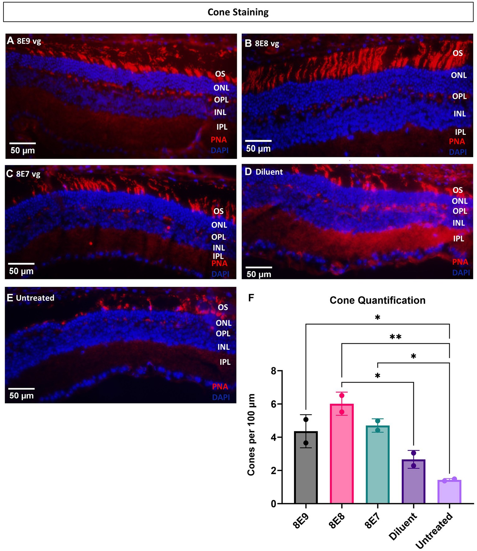
Frontiers The dose-response relationship of subretinal gene therapy with rAAV2tYF-CB-hRS1 in a mouse model of X-linked retinoschisis

All Protocols and Video Articles in JoVE
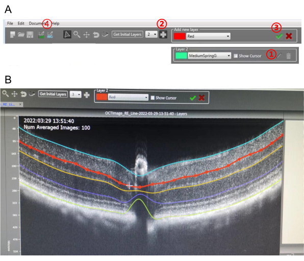
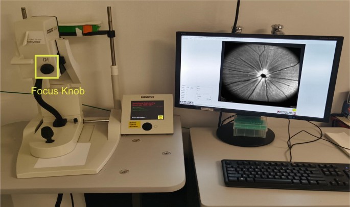
Image-Guided Optical Coherence Tomography to Assess Structural Changes in Rodent Retinas

Image-Guided Optical Coherence Tomography to Assess Structural Changes in Rodent Retinas
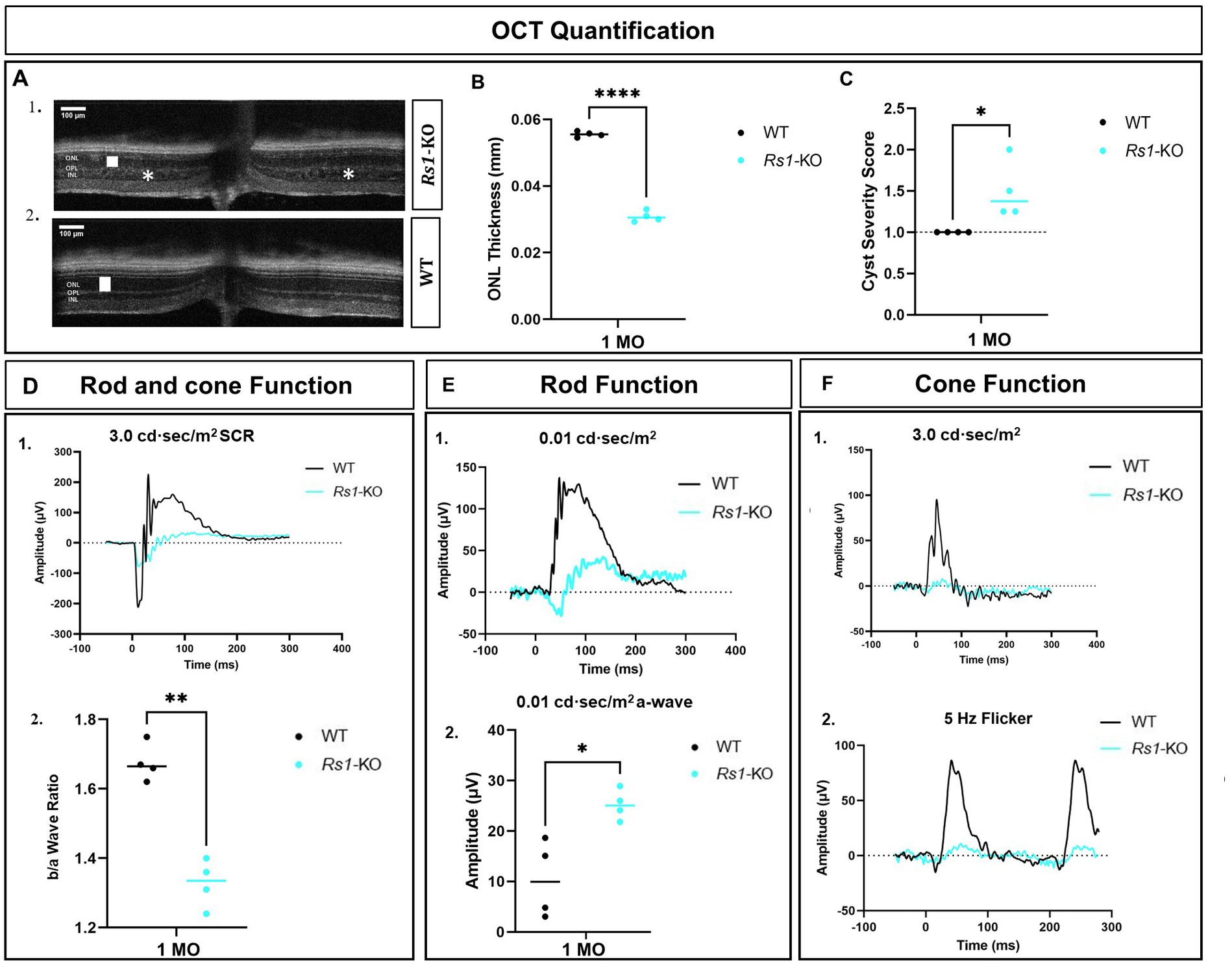
Frontiers The dose-response relationship of subretinal gene therapy with rAAV2tYF-CB-hRS1 in a mouse model of X-linked retinoschisis
Volker BÄCKER, Engineer, Dipl. Inform., French Institute of Health and Medical Research, Inserm, Biocampus Montpellier - US9

Emmanuelle SARZI, Professor (Assistant), Claude Bernard University Lyon 1, Villeurbanne, UCBL, Institut NeuroMyogène

Emmanuelle SARZI, Professor (Assistant), Claude Bernard University Lyon 1, Villeurbanne, UCBL, Institut NeuroMyogène

Optical Coherence Tomography: Imaging Mouse Retinal Ganglion Cells In Vivo
Optical Coherence Tomography: Imaging Visual System Structures in Mice

Topical Nerve Growth Factor (NGF) restores electrophysiological alterations in the Ins2Akita mouse model of diabetic retinopathy - ScienceDirect
Frontiers Early Retinal Defects in Fmr1−/y Mice: Toward a Critical Role of Visual Dys-Sensitivity in the Fragile X Syndrome Phenotype?

All Protocols and Video Articles in JoVE