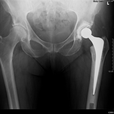
Total hip replacement, Radiology Case
This represents a case of total hip replacement. The patient presented to the emergency department with worsening pain in the left hip, groin, and leg. Imaging workup demonstrated effacement of the

Fig. 2.6, [In this case, the patient,]. - Personalized Hip and Knee Joint Replacement - NCBI Bookshelf

Revision Total Hip Arthroplasty using a Direct Anterior Approach in a Patient with Arthrogryposis Multiplex Congenita: A Case Report

Postoperative radiograph of the hip arthroplasty: what the radiologist should know, Insights into Imaging

Imaging of Total Joint Replacement

Dislocated hip prosthesis, Radiology Case

Wearing Out of Total Hip Replacement Surgery
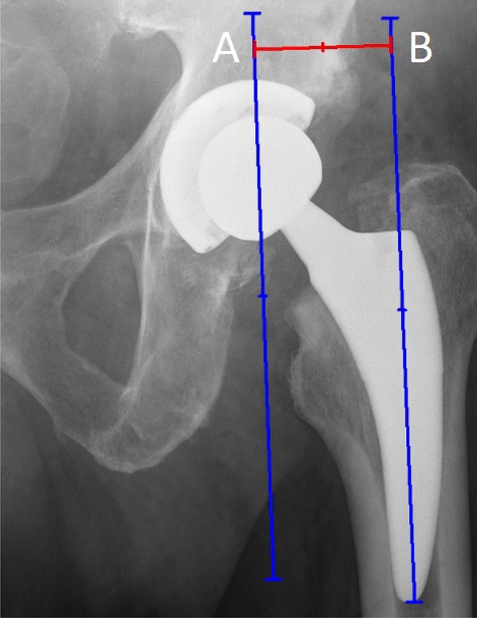
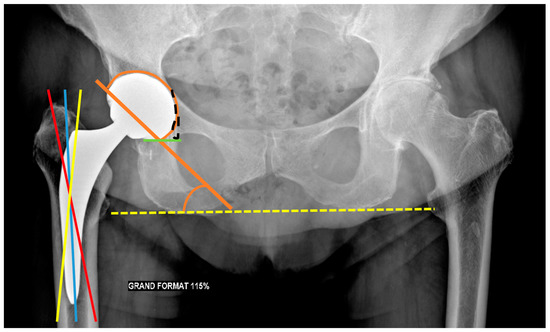
How to analyze postoperative radiographs after total hip replacement

JCM, Free Full-Text

Typical case 1. Male, 41 years old, right total hip arthroplasty. (A)
JCM, Free Full-Text

Incidence Of Heterotopic Ossification In Direct Anterior, 59% OFF