Calcification and mass abnormalities in breast mammogram scans
Download scientific diagram | Calcification and mass abnormalities in breast mammogram scans. The calcification distribution depicts tiny flecks of calcium as small white regions on the left side, while the mass is shown as a smooth, well-defined border on the right side. from publication: Multi-Graph Convolutional Neural Network for Breast Cancer Multi-Task Classification | Mammography is a popular diagnostic imaging procedure for detecting breast cancer at an early stage. Various deep learning (DL) approaches to breast cancer detection incur high costs and are prone to classify incorrectly. Therefore, they are not sufficiently reliable to | Breast Cancer, Convolution and Classification | ResearchGate, the professional network for scientists.
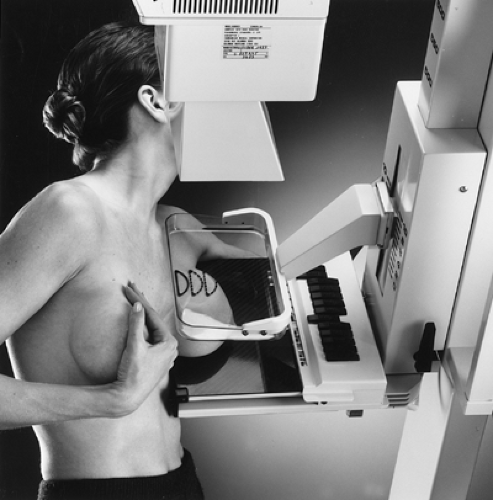
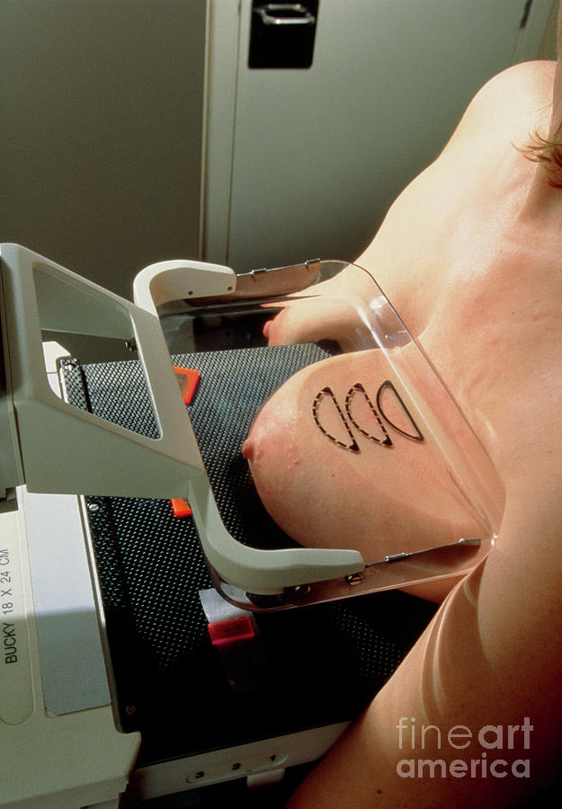
Atlas of breast cancer early detection

Calcification and mass abnormalities in breast mammogram scans

a) The cropping breast profile image of mdb111 for left MLO

Contrast enhanced mammography: focus on frequently encountered benign and malignant diagnoses, Cancer Imaging
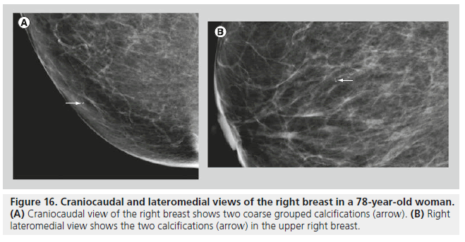
Mammography of breast calcifications

Brendan JENNINGS, Head of Graduate Studies
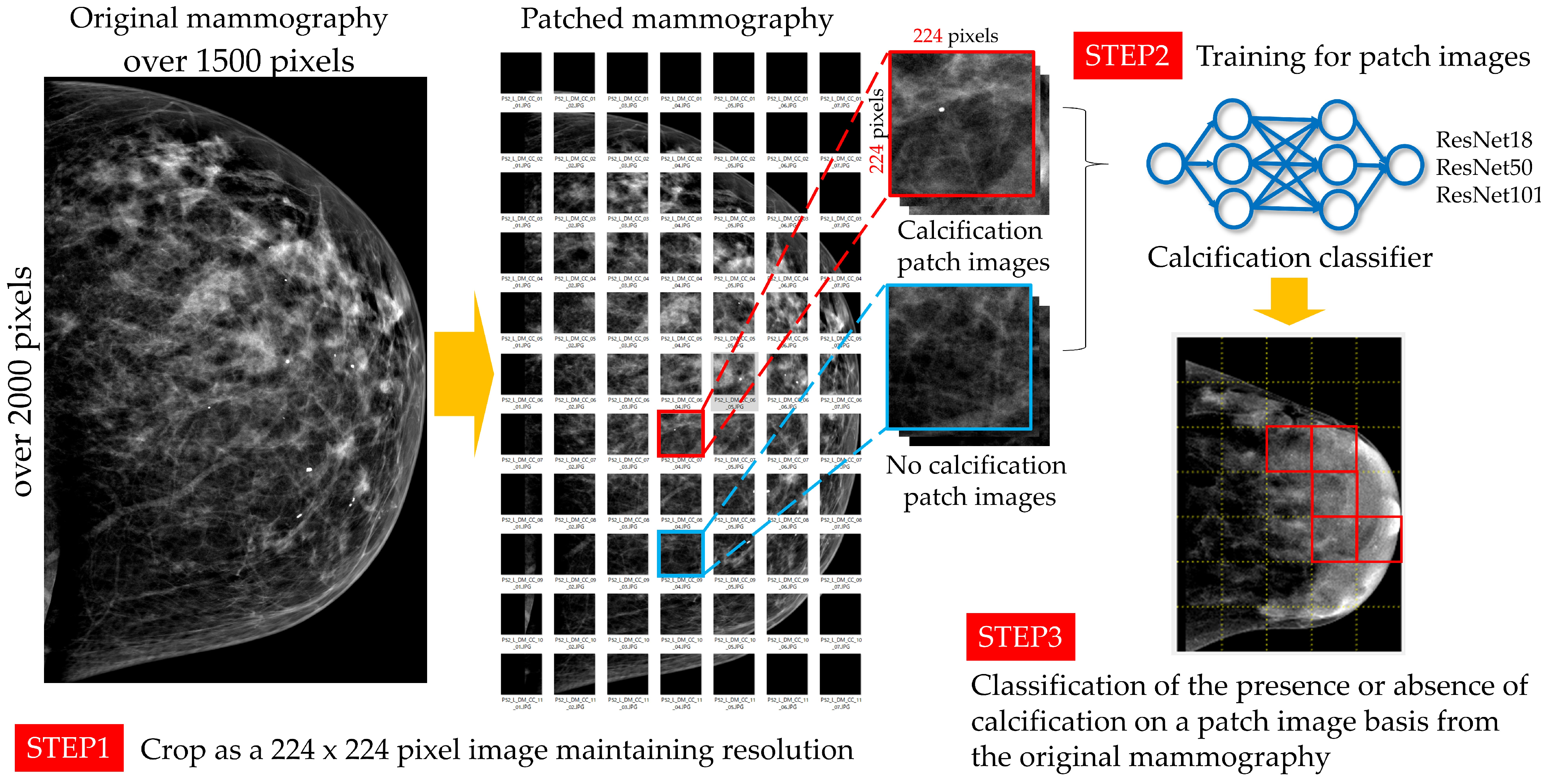
Algorithms, Free Full-Text

Use of Low-dose Chest CT Scan in the Evaluation of Breast Composition According to the Recommendations of BI-RADS Atlas-Fifth Edition, IJ Radiology
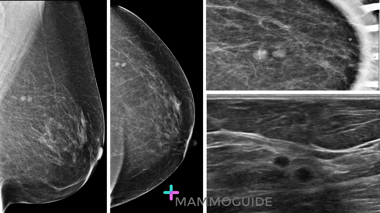
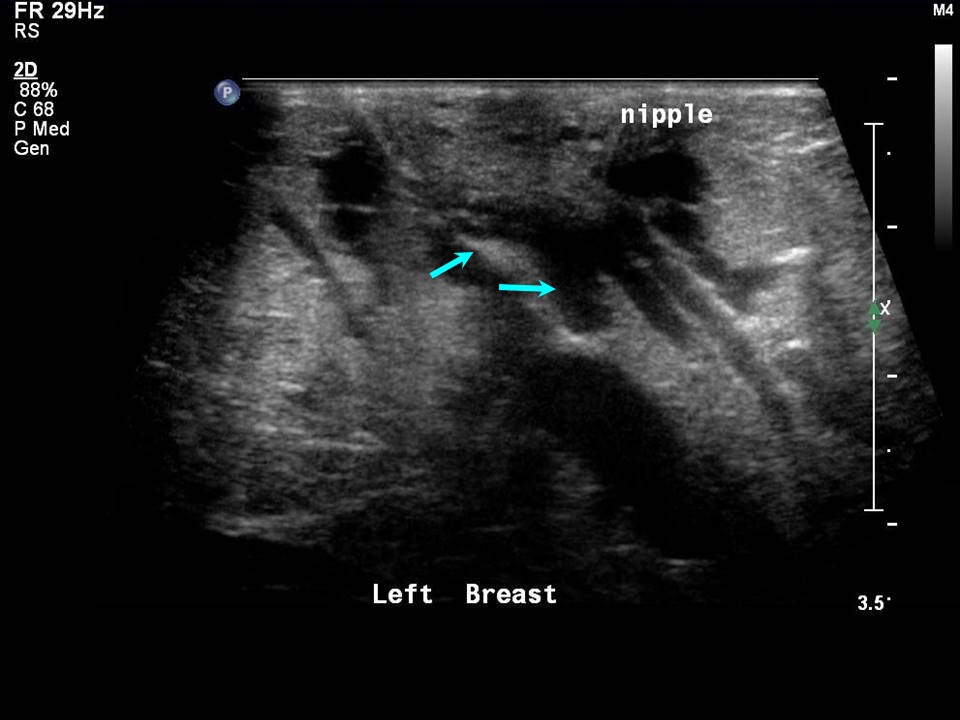
Breast Imaging Cases - MAMMOGUIDE - Learn Breast Imaging

PDF) Multi-Graph Convolutional Neural Network for Breast Cancer
Comparison of the quality of segmentation based on the number of
:max_bytes(150000):strip_icc()/breast-cancer-tumors-what-are-they-430277-v12-d91aad27f20b4f06aae6afc5a55868da.png)
Breast Masses: Cancerous Tumor or Benign Lump?

Example breast mammogram images with calcification and a mass
:max_bytes(150000):strip_icc()/430283_color-5bb3d89946e0fb00261df155.png)