Red and white blood cells in clot, SEM - Stock Image - C045/8688 - Science Photo Library
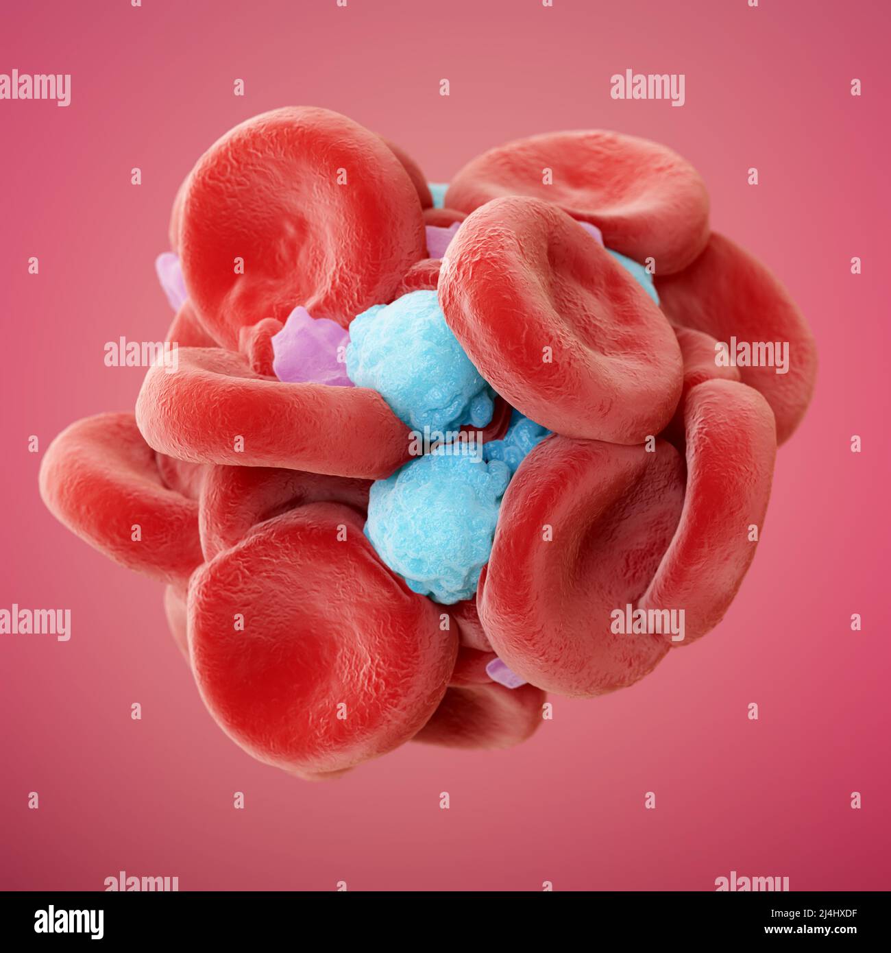
Red blood cells (erythrocytes) and a single white blood cell (leucocyte or leukocyte) in a fibrin mesh, coloured scanning electron micrograph (SEM). Formation of a blood clot with many erythrocytes (red) and a single leukocyte (white/blue) becoming entangled in a fibrin mesh (light brown). ANNE WESTON, FRANCIS CRICK INSTITUTE/SCIENCE PHOTO LIBRARY

Human Red Blood Cells Sciencephotography.com

Science Photo Library (@sciencephotolibrary) posted on Instagram

Red blood cells, white blood cells and platelets, SEM - Stock

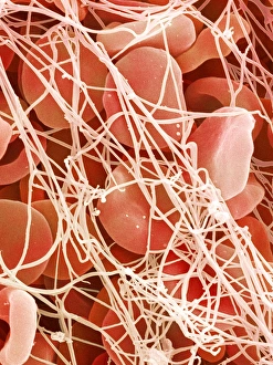
Blood Clot #3 by Secchi-lecaque/roussel-uclaf/col. V. Gremet/cnri
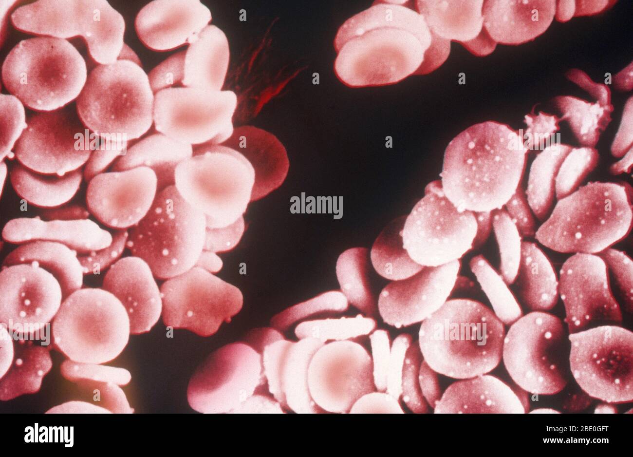
Sem red blood cells human hi-res stock photography and images - Alamy

Science Photo Library (@sciencephotolibrary) posted on Instagram
Blood Clot, Sem #2 by Steve Gschmeissner
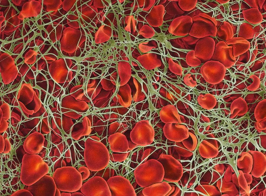
Blood clot. Coloured scanning electron micrograph (SEM) of a blood clot. The red blood cells (erythrocytes) are trapped in filaments of fibrin protein
Prints of Blood clot, SEM P260 / 0123

Blood Clot - Stock Image - C008/5083 - Science Photo Library

Blood clot, SEM - Stock Image - C056/3890 - Science Photo Library
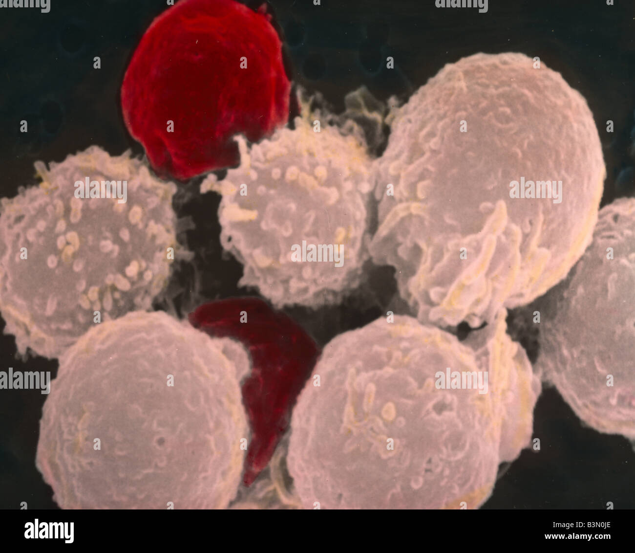
Human Red And White Blood Cells #1 Photograph by Dennis Kunkel Microscopy/science Photo Library
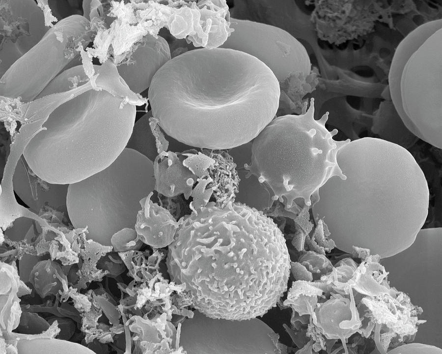
Human Red And White Blood Cells #1 by Dennis Kunkel Microscopy/science Photo Library
Fibrin red white blood hi-res stock photography and images - Alamy

Blood Clot, Sem #26 by Steve Gschmeissner
Red and white blood cell sem hi-res stock photography and images