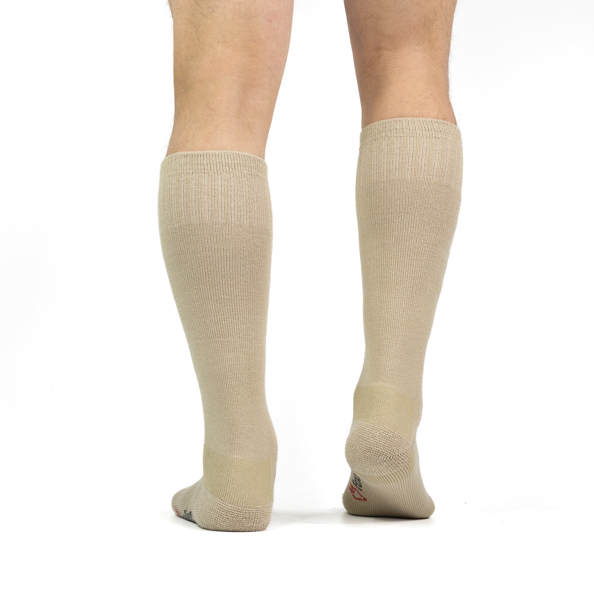
Comparison of MRI slices at the mid calf showing the anatomy with an IP

PDF) Relationship between medical compression and intramuscular pressure as an explanation of a compression paradox
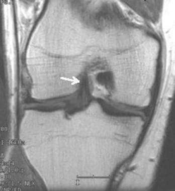
Anterior Cruciate Ligament (ACL) MRI: Practice Essentials, Anatomy, Mechanism of Injury
Brain MRI ATLAS

Fasciae of the musculoskeletal system: MRI findings in trauma, infection and neoplastic diseases, Insights into Imaging

White Matter Signals Reflect Information Transmission Between Brain Regions During Seizures

IJSPT V17N7 by IJSPT - Issuu

Jean-François UHL, Unesco Chair of digital anatomy, MD, FacPh, Paris Descartes, CPSC, Paris, Paris 5, UFR biological (Saints-Pères)

André CORNU-THÉNARD, Vascular Medical Doctor, Research in Compressiontherapy and Treatment of Elephantiasis by Reduction, Cornu-Thenard MD, FACPh -, Doctor André Cornu-Thenard; France, Paris, 75011 Faidherbe Street 2

Thoracic spine protocol (MRI), Radiology Reference Article

Neuromorphic Optical Data Storage Enabled by Nanophotonics: A Perspective

Fitzpatrick skin type and patterns of melasma

Comparison of MRI slices at the mid calf showing the anatomy with an IP
MIDA: A Multimodal Imaging-Based Detailed Anatomical Model of the Human Head and Neck

Presence of a valve at the saphenofemoral junction.