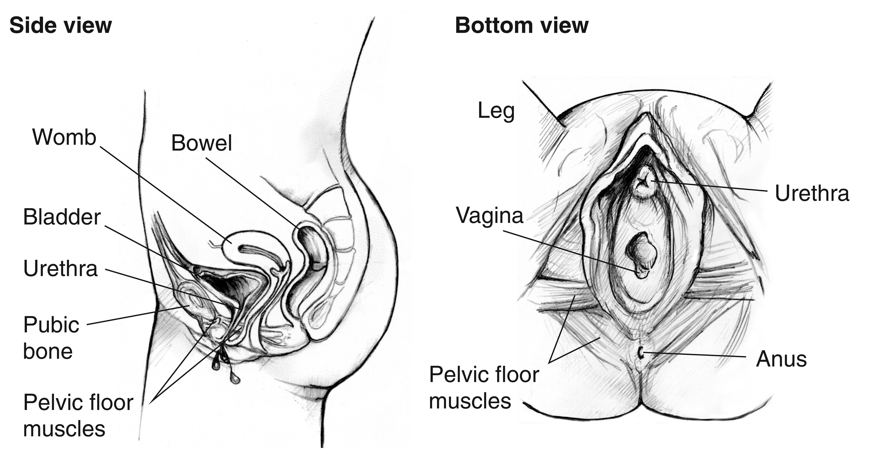
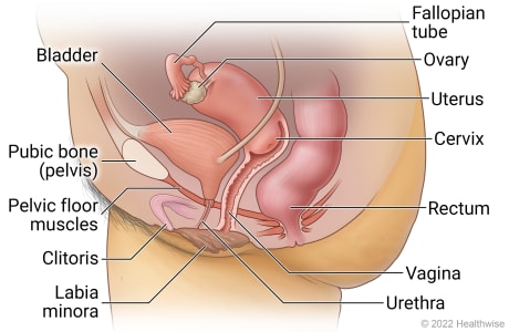
Side and bottom views of the female urinary tract - Media Asset
Two anatomical drawings of the female urinary tract. Drawing on the left is a side view with labels pointing to the womb, bladder, urethra, public bone, and pelvic floor muscles. Drawing on the right is a bottom view with labels pointing to the leg, ureth
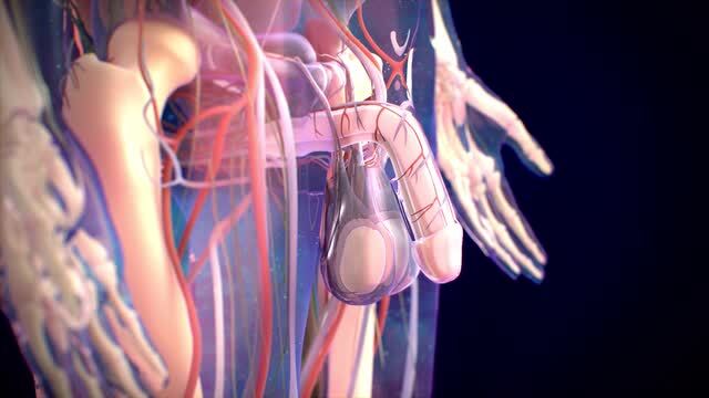
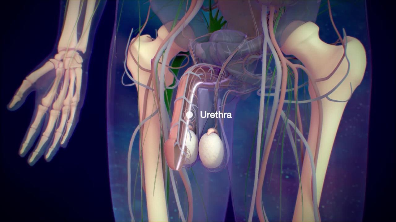
Structure of the Male Reproductive System - Men's Health Issues - MSD Manual Consumer Version
Pelvic exenteration: Radical but positive for carefully selected patients with pelvic cancer - Mayo Clinic

Human reproductive system, Definition, Diagram & Facts
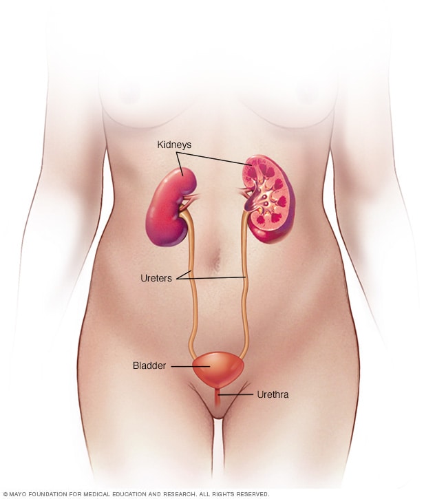
Urinary system: Organs, anatomy and clinical notes

Seven body organs you can live without
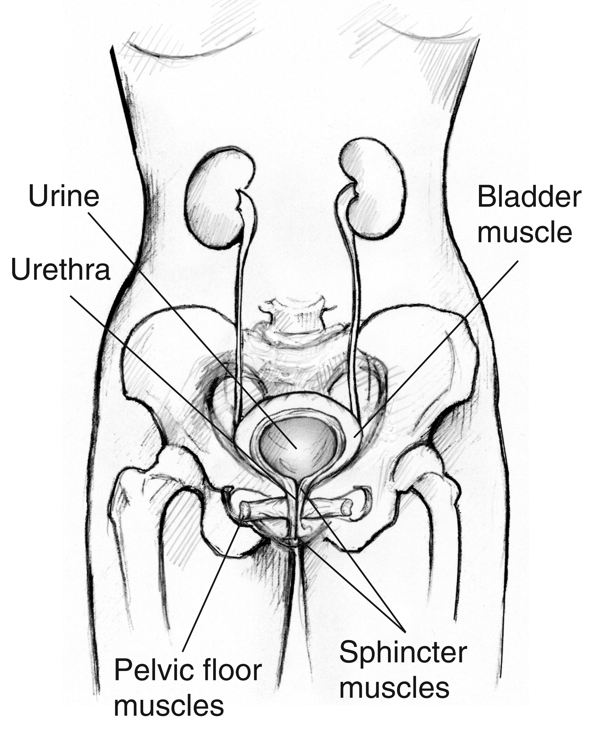
The front view of the female urinary tract. Labels point to pelvic floor muscles, sphincter muscles, bladder muscle, urethra, and urine - Media Asset - NIDDK
Bladder removal surgery (cystectomy) - Mayo Clinic
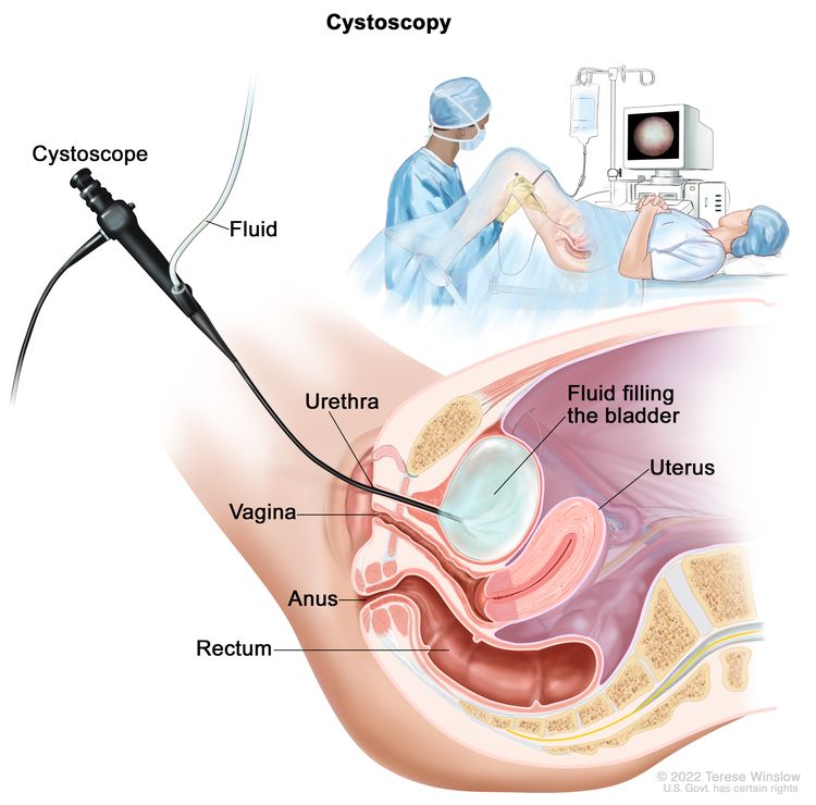
Urethritis - Kidney and Urinary Tract Disorders - MSD Manual Consumer Version
/images/vimeo_thumbnails/258819787/FRz0jezX7I2tJUyB5eQ_overlay.jpg)
Urinary bladder: Anatomy, function and clinical notes
Prostate Cancer – Early-Stage: Symptoms, Diagnosis & Treatment - Urology Care Foundation
Bladder Cancer Diagnosis - NCI

The Female Anatomy 101: An Intimate Look At Our Bodies - Axia Women's Health
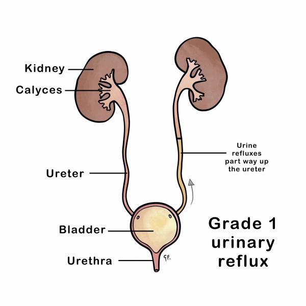
Micturating Cysto-Urethrogram (MCU)
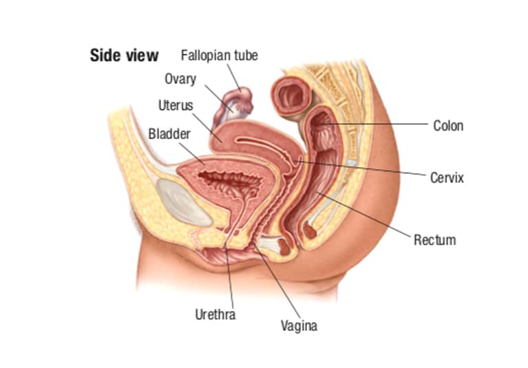
Side view diagram of the female urinary tract - Media Asset - NIDDK
:max_bytes(150000):strip_icc()/GettyImages-636080674-1f723ae1e1a7497dbdb38b2dcb132736.jpg)