Grey scale imaging (ultrasound), Radiology Reference Article
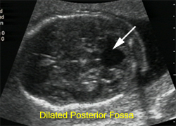
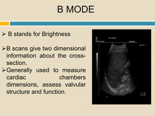
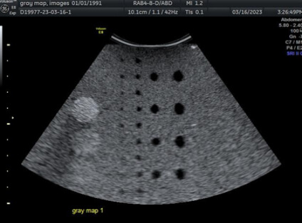
Commonly referred to as B (brightness) mode, the use of grey scale imaging in ultrasound renders a two-dimensional image in which the organs and tissues of interest are depicted as points of v

Grayscale ultrasound evaluation
CT of Calcifying Jaw Bone Diseases
50 More Shades of Gray: About Gray Maps - Ultra Select Medical
Review of X-ray and computed tomography scan findings with a promising role of point of care ultrasound in COVID-19 pandemic

ULTRASOUND CORNER: RANGE AMBIGUITY ARTIFACT - O'Brien - 2001 - Veterinary Radiology & Ultrasound - Wiley Online Library

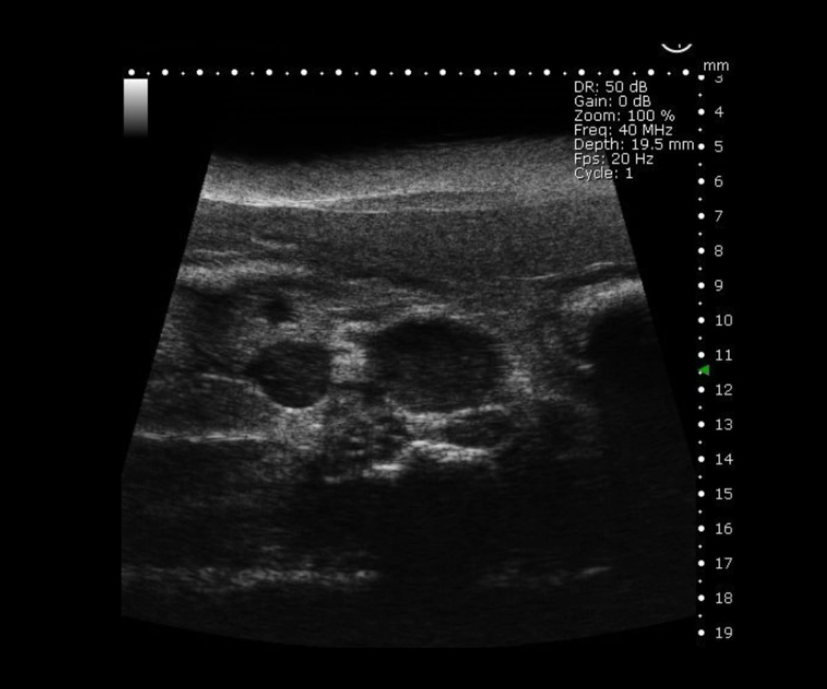
Automatic classification of ultrasound thyroids images using vision transformers and generative adversarial networks - ScienceDirect

TTG Imaging Solutions on LinkedIn: Routine chest CT often reveals patients at risk for cardiovascular…

To explore the pathogenesis of Bell's palsy using diffusion tensor image
Imaging features of breast cancer subtypes on contrast enhanced ultrasound: a feasibility study - ecancer

Role of gray-scale and color Doppler ultrasound in women with chronic pelvic pain Abdullah MS, Mousa WA, Ghobashy MA - Menoufia Med J

PDF] From Grey Scale B-Mode to Elastosonography: Multimodal Ultrasound Imaging in Meningioma Surgery—Pictorial Essay and Literature Review