Grey scale imaging (ultrasound) Radiology Reference Article
Commonly referred to as B (brightness) mode, the use of grey scale imaging in ultrasound renders a two-dimensional image in which the organs and tissues of interest are depicted as points of v
Diaphragmatic hernia - Radiology at St. Vincent's University Hospital

Orthoroentgenogram Radiology imaging, Prosthetic leg, Body anatomy

File:Radiopedia MRI brain metastasis - Copy.jpg - wikidoc
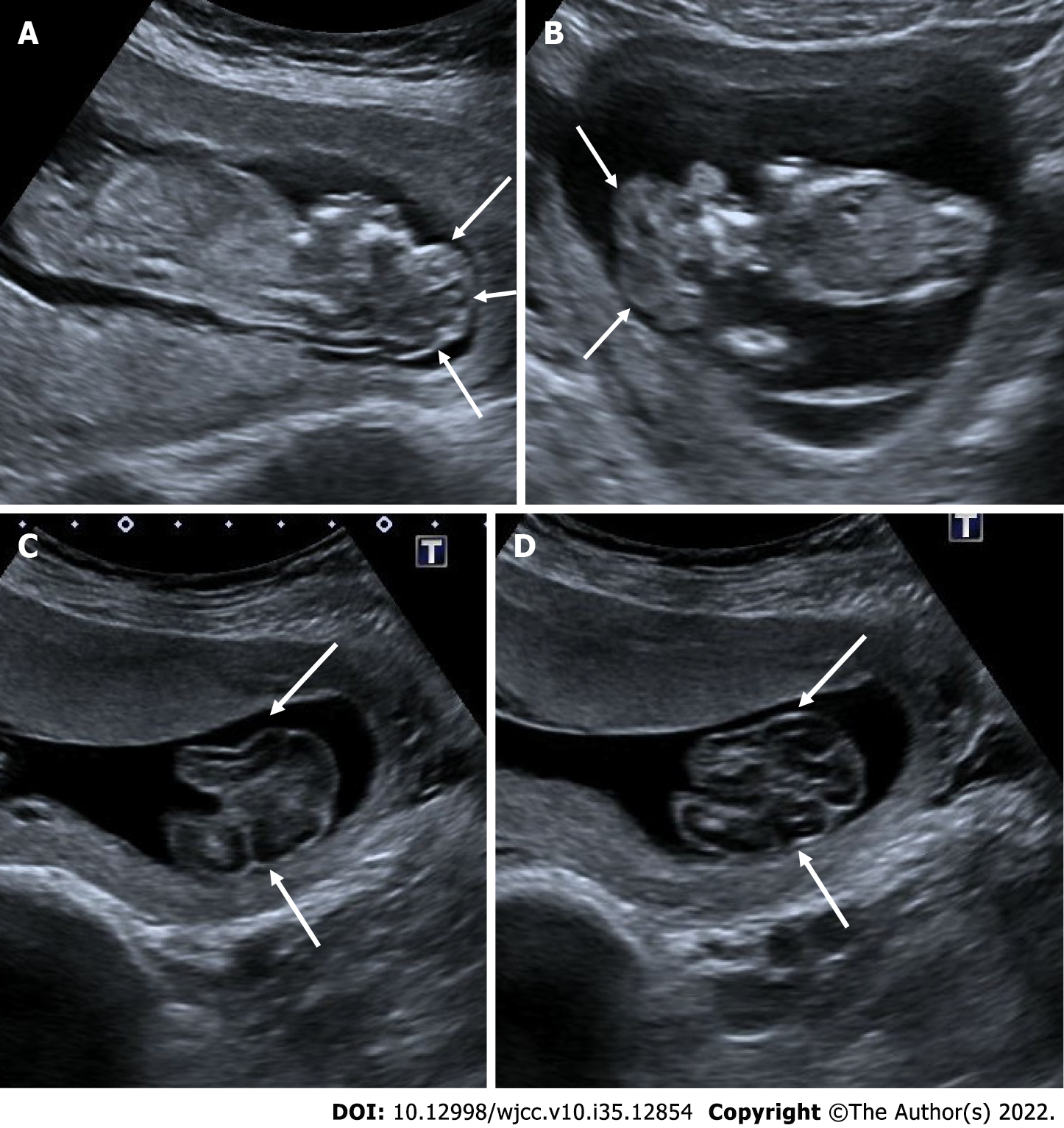
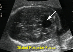
Antenatal imaging: A pictorial review

256 Shades of Gray – Explanation of Grayscale –

Antenatal imaging: A pictorial review

CNSeminars – Clinical Neuroanatomy Seminars on X: 🚀 #Neuroccino: #MedSAM - a game-changer in #medical image #segmentation! Trained on 1.57M images across 10 modalities, it excels in accuracy & versatility. A giant
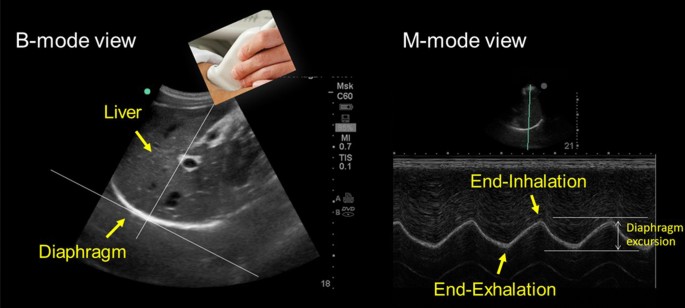
Internal Medicine Point of Care Ultrasound - IMPoCUS

Figure, Ultrasound: Gray scale images of] - StatPearls - NCBI Bookshelf

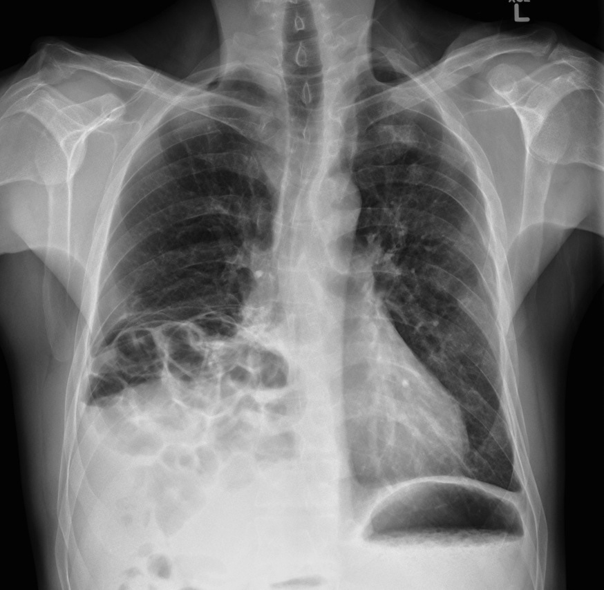
How does normal lung x-ray look?