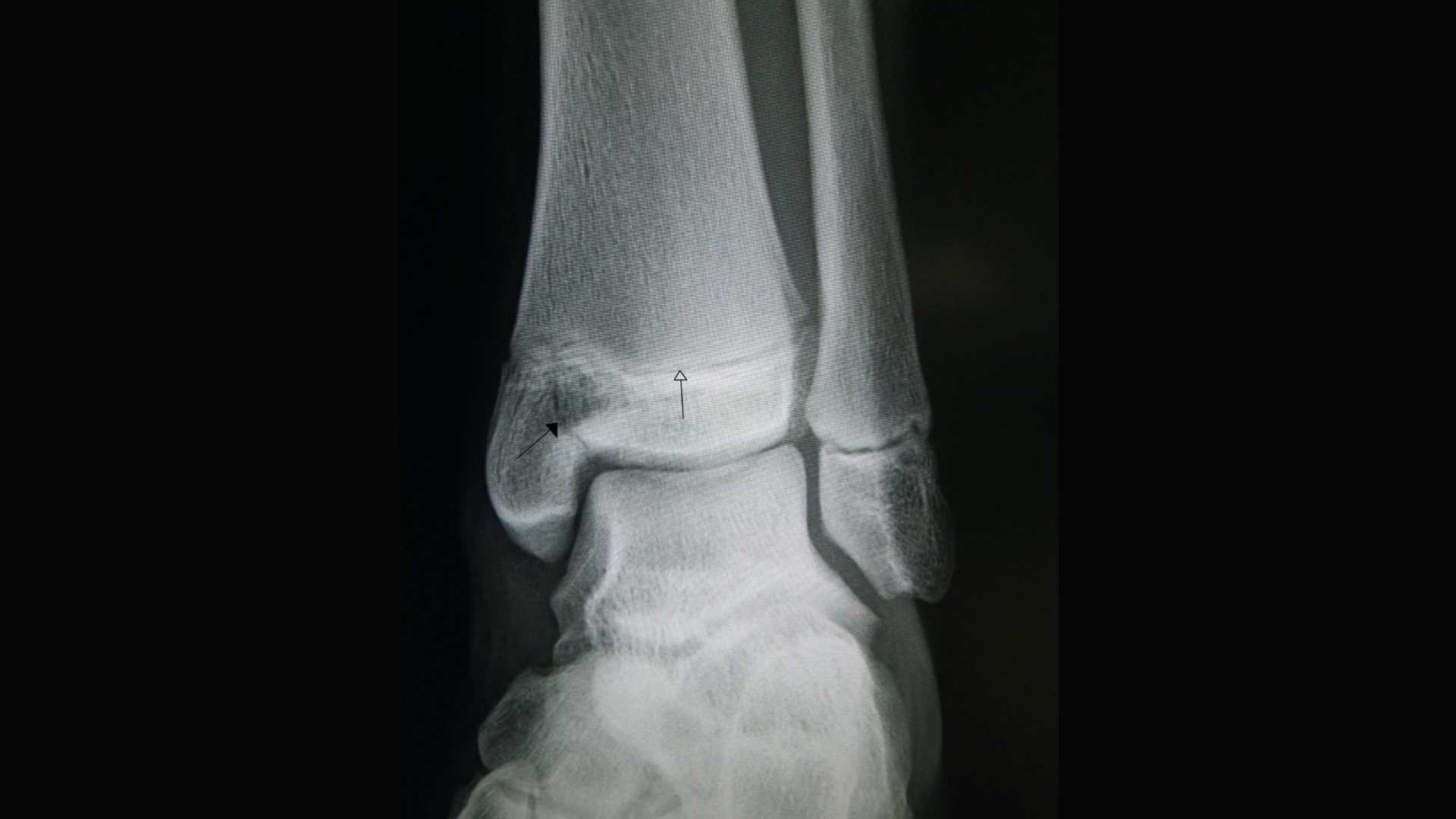
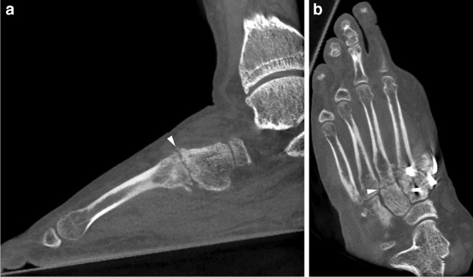
Foot X-ray of a 10 year-old male patient (white arrow indicates

25: Magnetic Resonance Imaging of Foot and Ankle Pathology
Musculoskeletal (MSK) X-ray Interpretation - OSCE Guide
Lower Extremity Plain Radiography (Chapter 2) - Clinical Emergency Radiology
Imaging of osteoarthritis from the ankle through the midfoot

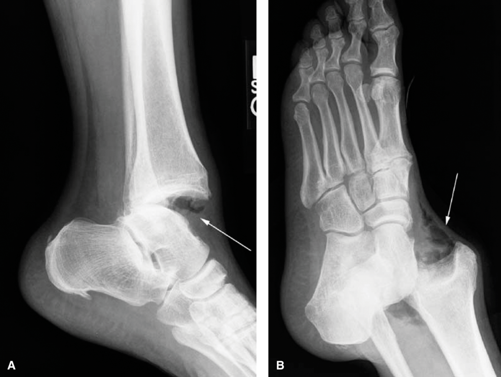
a) Lateral X-ray of the patient. (b) Red line (*) denotes a fracture

Extent of scrotal injury at examination under anaesthetic.

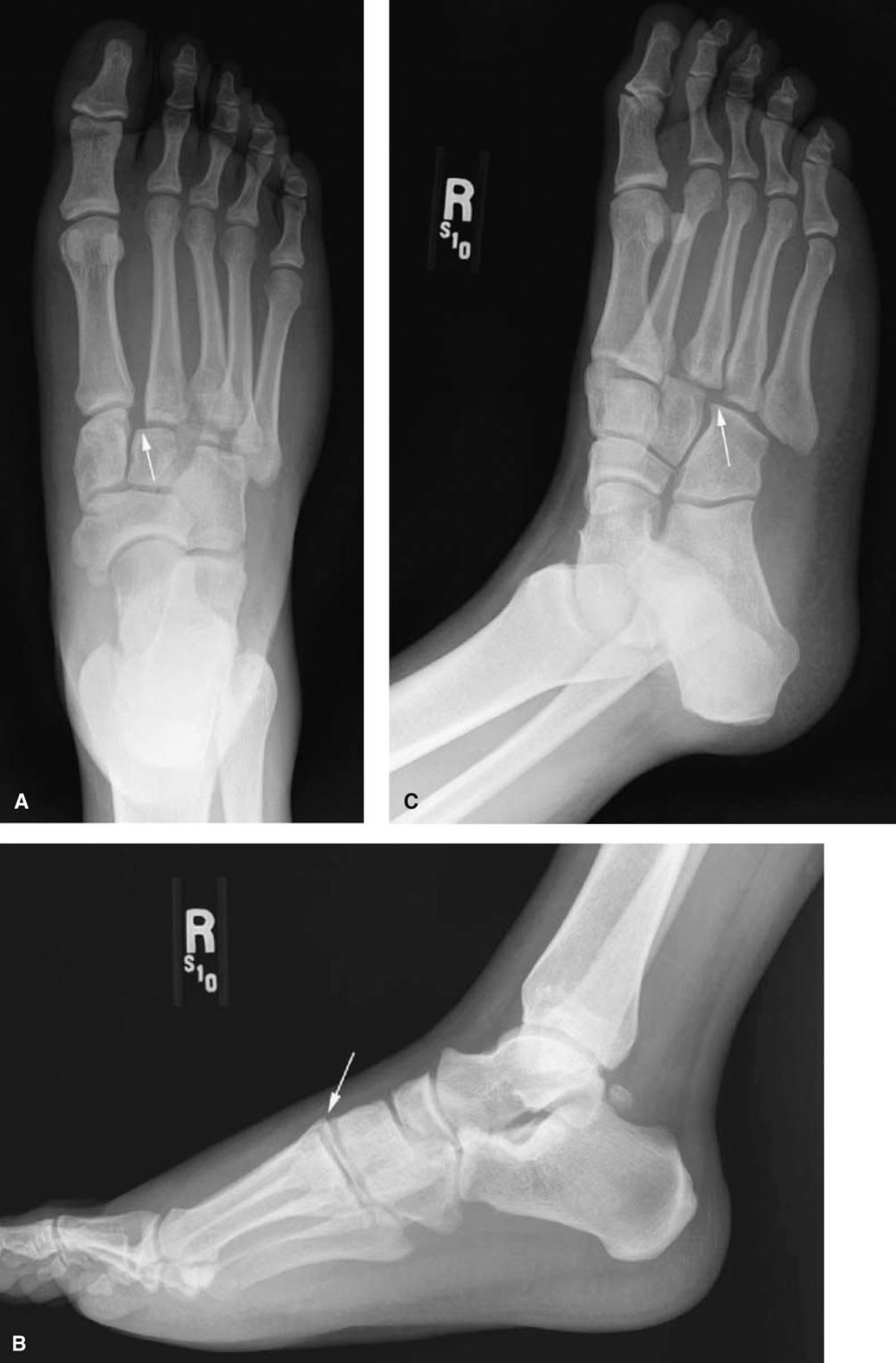
Acute Fractures and Dislocations of the Ankle and Foot in Children
Lower Extremity Plain Radiography (Chapter 2) - Clinical Emergency Radiology

PDF) Sprain Injury in a Child: Where is the Fracture Line?

Soyuz Drop Test Platform [24] Figure 3-9: Seat Testing at Various

X ray of the left foot of a 29 years old male patient. Fracture of the first metatarsal bone, Stock Photo, Picture And Rights Managed Image. Pic. BSI-BSIP-013095-064

EMRad: Radiologic Approach to the Traumatic Foot X-ray

Ozlem Bilir's research works Recep Tayyip Erdoğan Üniversitesi, Rize and other places

Fig. 3.1, a Chest X-ray showing cavitary lung lesions (white arrow) and upper lobe opacities (smaller red arrows) in 46 year old male. b Chest X-ray with the classic 'scattered millet seed
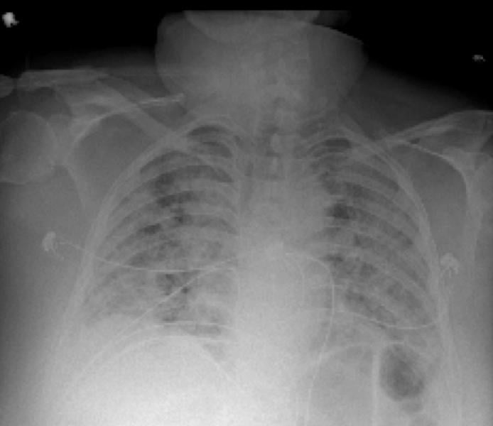
PHOTO GALLERY: How COVID-19 Appears on Medical Imaging